Post 6: The Three-Story Brain
Your brain is modular, comprised of three separate levels that each must separately learn that danger has passed.

Series II — Architecture of Mind
It is September 13, 1848. A railway construction crew in Vermont is blasting rock for the Rutland and Burlington Railroad. Phineas Gage, age twenty-five, is the site foreman. By all accounts he is an exceptional worker, reliable and well-regarded, with the kind of practical intelligence and judgment that have helped his bosses trust him to lead.
He is tamping black powder into a borehole when the charge fires prematurely. A thirteen-pound four foot long iron rod shoots upward through his skull. It enters beneath his left cheekbone, passes behind his left eye, and exits through the top of his head, landing some thirty feet away.
Remarkably, Gage survives. He is alert and talking within minutes. He walks, with help, to the cart that carries him to the nearest town. His physician, John Harlow, documents with careful amazement that the patient never loses consciousness. Within two months, Gage is physically recovered.
Though he lived, it is also true that Gage died that day. The person his friends and colleagues had known and relied upon, who they found steady, practical and trustworthy, was simply gone. In his place was someone who wore the same face and carried the same memories but could not be reached in the same way. His friends experienced a loss the medicine of the time had no language for: not the loss of death, which at least has its own completion, but something stranger and more disorienting.
His friends put it plainly: "Gage is no longer Gage."
In the wake of the accident he has become quite impulsive, easily frustrated, unable to make and keep plans. He does not lose his intelligence. He can reason, hold a conversation, and recall the past. But the coherence that Gage's friends once relied on, the sense that a steady person lived behind his eyes, is no longer there. Gage spends the rest of his life unable to maintain employment, drifting from job to job, eventually exhibiting himself alongside his iron rod at Barnum's American Museum in New York.
Mid-19th century medicine had no framework to explain what had happened to Phineas Gage. What kind of organ can be partially destroyed and produce something as specific as loss of selfhood while leaving speech, memory, and movement intact? What does this say about how the brain is organized? And if the brain is organized in a way that allows for this kind of targeted disruption, what does that mean for how the brain breaks down and how it can heal? These questions set in motion many years of accumulating scientific investigation that would ultimately provide answers. The same questions also, I would argue, sit beneath and have motivated all of the research I will lay out across this collection of posts which comprise Series I - Architecture of Mind.
Quick Guide: What You're About to Read
This post traces a line of scientific investigation that began with Phineas Gage in 1848 and continues to the present day. Along the way you will see how a series of pivotal discoveries (some accidental, some deliberate) gradually revealed the brain to be a modular system whose parts must coordinate to produce a coherent self. You will see what that architecture, once understood, tells us about how trauma fragments the brain's organization and how healing restores it.
Throughout this post, I will be offering two structural metaphors to assist your comprehension of the material. The first metaphor involves a three story building. The second metaphor involves an orchestra. Both metaphors are introduced in the main text when needed.
When you finish reading this post you'll understand:
- Why Gage lost himself while keeping his memory and speech, and what that tells us about brain organization
- The three functional regions: Brainstem, Limbic System, Cortex
- How these regions talk to each other, and what happens when that conversation breaks down
- Why Paul MacLean's 1960s synthesis was brilliant and largely correct, and where it went wrong in a way that matters for healing
Read time: 50–65 minutes total | 15–20 minutes for core concepts only
Difficulty: Medium: neuroscience concepts explained through metaphor and story
Heavy content: No. This is foundational architecture. The clinical illustration is non-violent.
Suggested stopping points: After the Coordination Principle section and its Pause Point if you want the essential framework; after the clinical illustration if you want the full picture.
Support Resources: If You Need Them
Neuroscience concepts can feel overwhelming. Remember: you are not trying to memorize the details of brain anatomy. Instead, you are learning a pattern.
The pattern you're learning: Your brain has different regions. They must communicate. Trauma disrupts that communication. Healing restores it.
Grounding technique: Before you read further, notice your feet on the floor. Take three breaths where the exhale is longer than the inhale. Feel that rhythm? That is your brainstem (your Rhythm Section) keeping time. It has been doing this since before you were born. You can trust it to keep the beat while you read.
The Investigation: How We Learned the Brain Is Organized (1848–1960s)
Phineas Gage's accident posed questions that mid-19th century medicine could not yet answer. In posing such questions, Gage's accident set in motion the scientific investigation of how the brain actually works. Results from this investigation accumulated slowly, however, and only cohered because other physicians appreciated the fundamental importance of the questions and were paying attention. Though there were a lot of studies that contributed to the accumulating knowledge, I here describe only the most important ones.
In 1861, thirteen years after Gage's accident, a French surgeon named Paul Broca treated a patient with a slow-growing tumor who had lost, over time, almost all ability to produce speech. He could understand what was said to him. He could think. But he could not select words to produce speech. When the patient died, Broca performed an autopsy and found the precise location of the patient's brain damage: in a specific region in the left frontal lobe, now called Broca's area.
That finding alone would have been significant. But what happened next clarified the picture tremendously.
In 1874, a German physician named Carl Wernicke described a series of patients who could produce speech but could no longer understand it. These patients talked, and they did so fluently, expressively, and at length. However, what they said was incoherent. They could not comprehend what was said to them. Upon autopsy, the brain damage in these patients was located in the left temporal lobe; a different part of the brain than where Broca's patient's brain damage was found.
Taken together, these two reports documented two groups of patients, each with a different kind of speech deficit, with each group having different damaged locations in their brains. From here, the connection between specific speech deficits and specific brain damage could be clearly seen. If one specific brain region is damaged, you lose speech production but keep comprehension. If you lose the other specific brain region, you lose speech comprehension but keep production. Broca and Wernicke had together demonstrated what neuroscientists today call a double dissociation. What it proved was this: language is not a unified capacity housed in a single place within the brain. Instead, language arises from the coordination of a system of brain modules. The brain was revealed to be an organized modular system.
The elegant double dissociation pattern revealed by Broca and Wernicke's work gave rise to a methodological insight that the following century's scientists built upon: you can learn what a particular brain region does by carefully studying what functions are lost when that region is damaged.
Researchers began gathering evidence through two pathways. Sometimes they observed focal damage after the fact, following accidental injury, stroke, or illness, then correlating the behavioral and functional changes they found with the location of the brain damage. In other cases they induced damage deliberately, creating a controlled lesion in a specific brain region, then studied what behavioral and functional changes followed.
The evidence accumulated slowly, different damage producing different deficits. That methodological pattern was repeated, study by study, across a century of careful observation.
By the 1950s and 1960s, enough data existed to enable neuroscientist Paul MacLean to organize it all into a coherent map. He identified three broad functional levels of the brain, gave them names, and showed how they fit together as an integrated system. He called it the triune brain. In MacLean's scheme, the brainstem managed survival and arousal, the limbic system processed emotion and social connection, and the cortex handled language, planning, and abstract thought. Information flowed between the levels, and the lower levels could sometimes override the higher ones under threat conditions.
MacLean's brilliant synthesis took 150 years of accumulated clinical observation and organized it into a framework that explained human behavior, predicted testable outcomes, and made intuitive sense of things like why emotions can overwhelm reason, or why trauma activates ancient survival instincts. MacLean deserves every bit of the credit that textbooks give him.
But MacLean did two different things, and they are worth separating. The first was to organize the evidence: the brain's modularity, its three levels, its hierarchical communication structures. MacLean got that part reasonably correct. But MacLean also proposed an evolutionary story, explaining how the brain built itself through sequential addition of new structures, with the reptilian brainstem having evolved first, followed by the mammalian limbic system, and crowned at last by the distinctly human cortex.
This evolutionary narrative was an interpretation, a 'just so' story to explain how the complex brain came to have its shape. This narrative was not required by the evidence MacLean had organized, which did correctly describe the brain's modularity. There was nothing in the data that required that the brain's modules had evolved in a particular sequence.
We have since learned that evolution does not generate and stack completely new layers each time an adaptation is required. It turns out fish have functional versions of all three brain regions. So do birds. So do reptiles. The brainstem, limbic structures, and cortex-like tissue were all present in early vertebrates more than 500 million years ago. What evolved across species was not the addition of new structures but rather that structures that were already there were elaborated upon under evolutionary pressure. The brain's cortex has been elaborated to an extraordinary degree in humans. In contrast, the brainstem we carry is a highly conserved system whose fundamental architecture has been retained across hundreds of millions of years of vertebrate evolution; a sign not of stagnation but of a design so well-suited to its function that evolutionary pressure has left it largely intact. Though the design is ancient, it is not crude.
Why does this correction matter for trauma and healing? Because the most common popular accounts of trauma recovery are built on a hierarchical model, whether they know it or not: calm your amygdala! get your prefrontal cortex back online! override your fight-or-flight response! The implicit framework is domination of the rational over the animal, the higher over the lower, the thinking brain conquering the feeling brain. This framing flows directly from MacLean's evolutionary story: if newer brain regions evolved because they better promoted survival, the older regions start to look like relics to be managed rather than partners to coordinate with and listen to.
The appeal of this framework was not accidental. MacLean's hierarchy mapped onto a much older tradition, one running from Descartes forward, that has long privileged reason over feeling, mind over body, thought over emotion. It also appeared to give biological grounding to Freud's theory of personality, with the id, ego, and superego mapping neatly onto the reptilian, limbic, and cortical brains. The hierarchy felt self-evident because it landed on cultural soil already prepared to receive it. MacLean's hierarchical account of the brain's architecture shaped how an entire generation of trauma clinicians thought about recovery.
The problem with this hierarchical model is not primarily that it misreads evolution. The problem is that it rests on a category error. The cortex's elaboration gave humans extraordinary capacities for language, planning, and abstract thought; all genuine survival advantages. But survival advantages are additive, not replacements. An addition to a building may increase its value, but it does not make the foundation obsolete. You cannot subtract the old in favor of the new and expect the structure to stand. The subcortical systems were not outmoded by the cortex's expansion any more than a building's plumbing becomes unnecessary because a penthouse is added above it. What changed with the cortex's elaboration was the range of what the whole system could do together, not the importance of the parts that were already there.
The subcortical systems, for example, the brainstem's arousal regulation and the limbic system's threat detection, are not inferior versions of what the cortex does. They are specialized for entirely different functions that the cortex cannot replicate. In their own domains, they operate faster, more automatically and more reliably than conscious thought. A cortex that attempts to dominate rather than coordinate with these subcortical regions tends to produce exactly the painful experiences trauma survivors know well: the exhausting gap between what you know and what you feel, and between understanding that you are safe and being unable to feel it.
What trauma disrupts is not a proper dominance hierarchy between brain regions but rather the coordination between them. And what healing restores is not the cortex's control over the brain's lower regions but the conversation between all three, where each level is addressed in its own terms, each taught in its own way that threat has passed. This way of thinking about the relationships between the brain's modules leads to a fundamentally different model of trauma recovery, and one that is better supported by the clinical evidence.
There is a philosophical implication running through all of this evidence that is worth naming. René Descartes, the 17th century French philosopher best known for the declaration cogito ergo sum, meaning "I think, therefore I am," proposed that mind and body are fundamentally different kinds of substance: the body material and extended in space, the mind immaterial and outside it entirely. This position, known as substance dualism, has shaped Western thinking about selfhood, soul, and consciousness ever since. The evidence accumulated across this post deals it a serious blow. If the mind were genuinely independent of its biological substrate, damaging the substrate should leave the mind untouched. But the cases we have reviewed show with increasing specificity that it does not: damage the prefrontal cortex and you lose the self, damage the hippocampus and you lose the ability to form new memories, sever the corpus callosum and you appear to produce two separate streams of awareness within a single skull. Substance dualism in its strong Cartesian form cannot easily survive this evidence. Whether something more remains, whether there is an irreducible subjective dimension to experience that physical description alone cannot capture, is a harder question to answer. Philosopher David Chalmers has influentially framed it as the hard problem of consciousness, and it remains a genuinely open question. But for the purposes of understanding trauma and healing, the evidence is clear enough: whatever consciousness ultimately is, its specific functions are tied with extraordinary precision to the hardware that houses them. Readers who want to follow this philosophical thread further can do so by reading the appendix note on consciousness and dualism at the end of this post.
Before we map the three levels of the brain, let's take a moment now to give you two metaphors I will use to help you envision the relationships between the brain's modules.
The first metaphor is a three story building, where the foundation provides stability enough for the upper floors to function, and where each floor can only do its work as it remains in contact with the other floors, above and below.
The second metaphor is of an orchestra, composed of a Rhythm Section at the base keeping fundamental time, a Melody giving the music its feeling, and a Conductor holding the score and trying to keep everything coordinated.
Neither metaphor is perfect. However, each in its own way may provide you a handle of sorts to better help you appreciate the pattern of relationships governing how the brain's modules are arranged. They do not offer an exact map of the brain, but having them at hand should make what follows considerably easier to see and hold.
What happened to Gage makes more sense when you consider the brain as a modular structure that was, in his case, partially damaged. Gage lost the part of the brain that helps the other parts of the brain to coordinate, specifically the region that integrates emotional information with deliberate planning, and that holds impulses in conversation with judgment. The orchestra metaphor will work best here: In Gage's damaged brain the Rhythm Section continued to keep the beat and the Melody kept playing. However, the Conductor's chair was now empty. And without Gage's brain retaining the ability to coordinate among its component parts, the coherent, integrated person his friends had known was simply gone.
Hold this corrected frame as we now map the brain's architecture itself.
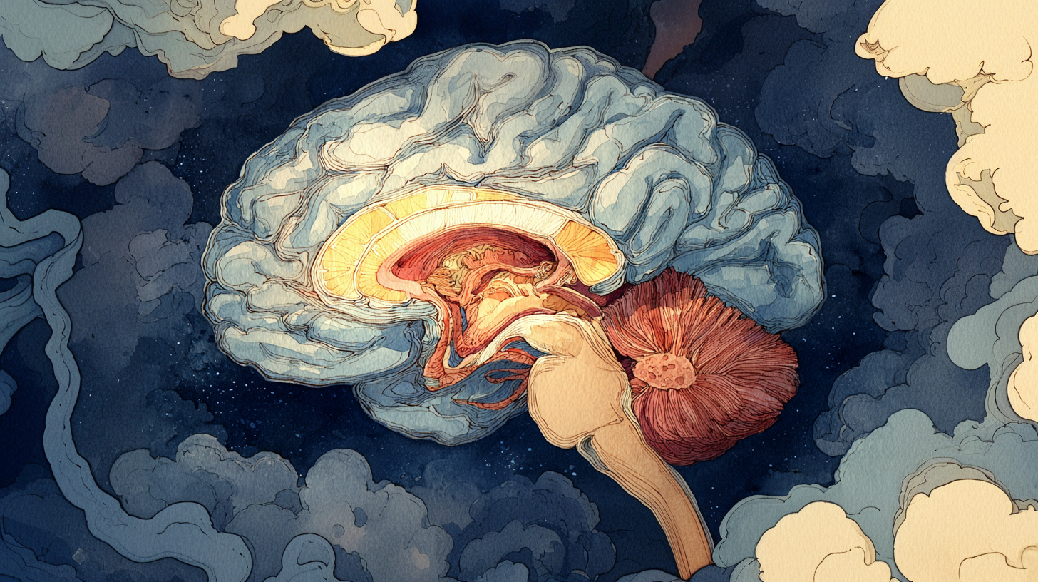
The Architecture: Three Integrated Levels
MacLean's synthesis produced a map of the brain's major divisions. It described three broad functional regions, each processing experience in its own distinct way, each serving a different purpose, all normally in constant communication with the others. In the following sections I will step you through each of these brain regions in more detail to give you a clearer sense of what each one does and how it contributes to the coordinated whole.
Level 1: The Brainstem: Your Survival Baseline
At the very core and bottom of your brain, where it connects to your spinal cord, lies your brainstem. The brainstem is a thick stalk running from the base of your skull down into your spine, roughly the diameter of your index finger. Everything else in your brain that makes you recognizably human is built around and on top of it.
The brainstem has been conserved largely unchanged across vertebrates for over 500 million years. If you imagine your brain as a three-story building, the brainstem is the foundation and the building's entire support infrastructure. Alternatively, if you imagine your brain as an orchestra, the brainstem corresponds to the Rhythm Section, the percussion and the bass, keeping fundamental time regardless of what the other sections might be doing.
The brainstem manages the most fundamental functions: breathing, heartbeat, sleep-wake cycles, reflexes, basic arousal. It does not think. It does not plan. It does not remember your childhood. It does one thing: it keeps you alive.
More specifically, your brainstem is responsible for arousal regulation. Think of a dimmer switch on a light. The brainstem controls that switch, shifting you between deep sleep, alert wakefulness and crisis activation. It reads your body's needs and the environment's demands and adjusts your arousal level to match. This happens automatically and entirely outside your awareness. You do not decide to breathe more shallowly when frightened; your brainstem causes it to occur. You do not choose what startle reflex to have when you hear a sudden noise; your brainstem executes that reflex within milliseconds.
The evidence for what the brainstem actually does comes, as with so much in neuroscience, from observing what is lost when it is damaged.
When a stroke destroys tissue in a specific region of the brainstem called the ventral pons, the result is something neurologists call locked-in syndrome, first popularized by Plum and Posner in 1966. The patient is fully conscious. Fully aware of their surroundings. Cognitively intact. But completely unable to move or speak, because the brainstem's motor pathways have been severed. The thinking brain is present. The emotional brain is present. But the foundation's wiring has been cut, and everything above it is stranded.
Brainstem damage can also reach the most basic act of all: breathing. Lesions in different parts of the brainstem disrupt breathing in different and specific ways, which tells us that different brainstem regions manage different aspects of respiratory control. Damage far enough down, toward a region called the medulla, can eliminate the automatic drive to breathe entirely; a condition known as Ondine's syndrome, in which a person can breathe voluntarily but will simply stop breathing the moment they fall asleep. The body no longer reminds itself to do what it has done since before birth.
And in 1949, the researchers Moruzzi and Magoun demonstrated through direct experimental work that the brainstem's reticular formation, a network of nuclei threaded through its core, is the primary source of arousal itself. Damage here does not merely reduce wakefulness. It extinguishes it.
Taken together, these findings confirm something important: the brainstem is not background infrastructure. It is the engine. Without it, nothing else runs.
Level 2: The Limbic System: Your Emotional and Relational Brain
Sitting above and around the brainstem, wrapping around it the way a second story might be built around an existing central pillar, is the limbic system. Its structures form a ring that partially encircles the top of the brainstem and extends upward into the protected interior of the brain, buried beneath the cortex.
If your brain is like a three story building, then the limbic system is a complex of interconnected rooms on the second story where your motivations (your emotions, drives and social bonds) all live. Alternatively, if your brain can be compared to an orchestra, the limbic system would correspond to the Melody, from which any music produced would get its feelings; the swells of joy, the minor keys of grief, the sharp staccato notes of fear or anger. This collection of interconnected structures evolved alongside the emergence of mammals from the broader family of vertebrates. Mammals were animals that did something new: they nursed their young, formed social bonds and experienced something recognizable as affection.
The limbic system includes the amygdala (threat detector), the hippocampus (long-term memory encoder), the hypothalamus (hormone regulator), and the cingulate cortex (emotional processor). These structures are not unique to humans. Other vertebrates have versions of them that function similarly, though they are organized differently. Birds, for instance, accomplish similar emotional and social processing through brain structures that look quite unlike the mammalian limbic system. The limbic architecture in other mammals, however, is recognizably similar to our own. Across all mammals, the limbic system is where motivation lives.
What does the limbic system actually do to produce motivation? Simply put, it continuously analyzes your situation and sets in motion instinctually prepared survival routines fitted to the situation it detects. In the time it takes you to consciously register that a situation is even happening, your limbic system has already evaluated it. A facial expression, a tone of voice, a body posture, a smell, shadow or light: before your thinking mind has formed an opinion, your emotional brain has already decided, its response already underway: safe or dangerous? accepting or rejecting? opportunity or threat?
We know the amygdala functions as a threat detector partly from a rare genetic condition called Urbach-Wiethe disease, which selectively damages it. People with this condition can think clearly, see faces, and recognize people intellectually, but they cannot feel fear in response to threatening situations. They know, in the cortical sense, that something is dangerous, but they do not feel afraid. The signal that would normally trigger fearful emotion simply is not generated.
We also know that the limbic system stores memories in emotional context; that you remember not just what happened but how it felt when it happened. We know this because of precisely the kind of double dissociation evidence described earlier. Two sets of patients, two different kinds of damage, two complementary losses that together reveal how the system is organized.
Studies of patients with hippocampal damage show us one half of the picture: demonstrating the brain's capacity to form emotional memories could remain after the capacity to form narrative memories had ceased. The most studied of these patients was Henry Molaison, known in the literature as H.M., whose medial temporal lobes, including most of his hippocampus, were surgically removed in 1953 to treat his epilepsy. After surgery he could no longer form new conscious memories. He could have a full conversation and moments later have no recollection of it having occurred. However, despite Molaison's inability to explicitly remember what had just happened to him, his capacity for emotional learning remained intact.
The capacity of amnestic patients like H.M., who could no longer form new explicit memories, to still be able to learn emotional content had been demonstrated years earlier in 1911 by Swiss psychologist Édouard Claparède, who concealed a pin in his hand when he shook hands with a patient with severe amnesia of a different origin. On subsequent meetings the patient refused to shake Claparède's hand but could not explain why, confabulating various reasons for their reluctance. Claparède's patient's ability to form new narrative memory was gone, but their ability to remember how they felt about shaking Claparède's hand, their wordless sense that something about Claparède required caution, remained.
Studies of patients with amygdala damage show us the other half of the picture: demonstrating that the brain's capacity to form narrative memories could remain after the capacity to form emotional memories had ceased. The clearest cases of this phenomena again came from Urbach-Wiethe disease. These patients can form and retrieve explicit memories of events normally: they remember what happened, when, and in what sequence. But their memories carry no emotional charge, the events they remember having been encoded without information about feeling. They recall the fact of an experience without any sense of what it meant.
Together these two patterns confirm what each alone could not: that the narrative and emotional dimensions of memory are stored through distinct but normally coordinated systems. When they work together, the full memory is encoded: both what happened and how it felt. When the coordination between the two memory systems breaks down, memories fragment and come apart. The normal coordination between the two memory systems explains why a particular song can drop you into the emotional atmosphere of a summer ten years ago before you have consciously recognized why your mood has changed. The feeling arrives before the story does, because they travel different routes.
The limbic system is also the neural home of social attachment. We know this not just from behavior but from what happens to the brain when social attachment has been disrupted. In the late 1950s, psychologist Harry Harlow began separating infant rhesus monkeys from their mothers, initially to prevent disease from spreading through his laboratory colony. What he had not anticipated was what the separation would do to the animals. Deprived of social contact during critical early periods of their development, the young monkeys developed profound and lasting interpersonal disturbances that damaged their ability to form normal social bonds. Instead of offering normal care-seeking behaviors, these young monkeys demonstrated self-directed repetitive behaviors and a kind of collapsed emotional life that witnesses found deeply disturbing. The monkeys had survived their early social deprivation but something essential inside them had failed to develop.
Decades later, a research team led by Lee Martin examined the brains of socially deprived monkeys and published their findings in the Journal of Neuroscience in 1991. They found measurable neurochemical changes: depleted levels of neurotransmitter markers in the striatum and basal ganglia, the circuits underlying motivation and reward. It turned out that the monkey's early life social deprivation had not just shaped their later-life behavior, but had also altered their brain chemistry in ways that persisted into adulthood.
The same pattern has been documented in human children. In 2001, a research team led by Harry Chugani used PET scans to study the brains of ten children who had been adopted from Romanian orphanages. These children had spent their earliest years living in institutions so severely under-resourced that meaningful caregiver relationships were essentially unavailable to them. The PET scans, which measure metabolic activity in living brain tissue, revealed reduced metabolic activity in limbic structures, suggesting less coordinated communication between them. The very circuits that would normally have supported these children's ability to regulate emotionally and seek social connection were less well coordinated than in the brains of children raised in ordinary family environments.
Separate neuroimaging studies, including a 2010 MRI study by Nim Tottenham and colleagues at Cornell studying 78 children, 38 of whom had previously been institutionalized, documented enlarged amygdala volume in children who had experienced prolonged orphanage care, with later adoption associated with larger amygdala volumes and greater difficulty regulating emotion. One plausible interpretation of this finding is that chronic social deprivation had pushed the brain's threat-detection system into a state of overdevelopment, as we might expect to find in a brain that had spent its earliest years adapting to an environment where frustration was more reliably present than satiation.
Taken together, these findings confirm something that is easy to state but not terribly obvious and therefore vitally important to appreciate: the brain's limbic system does not develop in isolation. It develops in relationship. The social bonds that the limbic system is built to support are also, in a very real sense, the very environment capable of building the limbic system. This is the neurobiology underlying John Bowlby's observation that human beings are born seeking connection. The seeking of social connection is wired in and supports its own construction.
A few things are worth keeping in mind about the limbic system as we move forward. It is where the brain's instinctual emotional and motivational systems are located. These systems operate largely automatically; they do not wait for you to make a decision, and you do not experience yourself making one when they activate. They are not rational in nature but survival-oriented, shaped by evolution to respond faster than thought allows. What this means in practice is that you cannot think your way into or out of limbic activation. You cannot reason with it, argue it down, or appeal to its better judgment. It speaks a different language than the cortex, and it responds to different kinds of input. To work with it, you have to meet it on its own terms.
Level 3: The Neocortex, Your Conscious, Planning, Language Brain
The neocortex is a convoluted blanket draped over the entire top of your brain, deeply folded so as to fit its enormous surface area inside your skull and enveloping all of the structures beneath it. The cortex (Latin for "bark," like the bark of a tree) is the thin outer layer of the structure, the gray matter visible when you look at pictures of the brain. All neocortical regions are interconnected by white matter connections carrying signals between each other and with the limbic and brainstem structures beneath them.
Right now, you are reading these words with your neocortex. This is the region that makes possible abstract thought, language, planning, imagined futures, remembered pasts, understanding other people's minds, and constructing narratives about why things happen.
If your brain is like that three-story building, then the neocortex is the upper story, housing the executive suite, the planning department, and the library. It is the place where decisions get made when there is time to think. Alternatively, if your brain is better compared to an orchestra, the neocortex is akin to the Conductor who holds the score, understands the lyrics, and tries to keep all the sections coordinated.
The neocortex is proportionally larger in humans than in most other animals, but it is not a fundamentally new structure. Our vertebrate ancestors already had a simpler cortical structure which became elaborated upon as humans evolved.
The manner in which the human neocortex has become elaborated is what makes it unique within the brain's architecture. It is not so much that it is "most advanced" or "best." What makes it special is its flexibility. The brainstem does the same thing every time, its reflexes operating automatically. The limbic system also does its work automatically, evaluating safety or threat within milliseconds. In contrast to these other structures, the neocortex can consider and reason. It can learn new patterns. It can imagine alternative futures. It can see how things fit together without those things being directly in view. It can question its first reaction. It can revise its own conclusions.
This flexibility is powerful. It is also expensive. The cortex requires massive energy to operate, and it operates much more slowly than the automatic systems below it. Under sufficient threat, the brain often partially takes the cortex offline to preserve energy for the survival systems. In such cases the Conductor goes quiet, but the orchestra continues to play.
The neocortex is itself a compound structure, organized into four broad regions. As throughout this post, we know what each region does because of what is lost when it is damaged.
At the rear of the brain sits the occipital lobe, dedicated almost entirely to processing vision. Damage here produces cortical blindness, a condition in which the eyes themselves are intact and functional but the person cannot see, because the brain can no longer interpret what the eyes are sending it. The visual world arrives but is never assembled.
Wrapping around the sides of the brain are the temporal lobes, handling hearing, aspects of memory, and language comprehension. This is where Wernicke's area lives, in the left temporal lobe, the region we encountered earlier in this post. Damage there, as Wernicke showed in 1874, produces fluent but incoherent speech: the ability to produce words without being able to understand them. The temporal lobes also contain regions specialized for recognizing faces. Damage to these areas produces what the German neurologist Joachim Bodamer named prosopagnosia in 1947, describing patients who had lost the ability to recognize familiar faces, including in some cases their own face in a mirror, while leaving all other visual processing intact.
At the top and toward the rear sit the parietal lobes, handling spatial processing, body awareness, and the integration of information from multiple senses. Damage to the right parietal cortex can produce one of the most disorienting deficits in neurology. In 1978, neurologists Edoardo Bisiach and Claudio Luzzatti described patients who systematically ignored everything on the left side of their world, not because they could not see it, but because the brain had stopped modeling that space as real. Patients with this condition, known as hemispatial neglect, eat only the food on the right side of their plate, draw only the right half of a clock face when asked to draw one, and are genuinely unaware that anything is missing.
At the front of the brain sit the frontal lobes, responsible for voluntary movement, planning, decision-making, and impulse regulation. The motor cortex, which controls voluntary movement, occupies the rear border of the frontal lobe. In a landmark series of surgeries beginning in the 1930s, neurosurgeon Wilder Penfield mapped the motor cortex precisely by applying tiny electrical stimulations to the cortical surface of patients who remained awake during epilepsy surgery. Each stimulation produced a specific movement in a specific body part, allowing Penfield to construct a detailed map showing which cortical region controls which part of the body. The resulting image, published with his colleague Edwin Boldrey in 1937 and refined through subsequent decades of work, is called the cortical homunculus, or "little man." It depicts a distorted human figure draped across the motor cortex, with the hands, face, and mouth enormously enlarged relative to the trunk and legs, reflecting the disproportionate amount of cortical territory devoted to fine motor control in those areas.
It is worth pausing here to note that the neocortex is divided into two hemispheres, left and right, each of which processes information from the opposite side of the body. The two hemispheres are connected by a thick bundle of nerve fibers called the corpus callosum, which allows them to coordinate and share information continuously. We know what the corpus callosum does because of what happens when it is surgically severed. In the 1960s, neuroscientists Roger Sperry and Michael Gazzaniga studied patients who had undergone this surgery as a treatment for severe epilepsy. When information was presented to only one hemisphere at a time, the two sides of the brain behaved as if they belonged to different people. Most strikingly, the left hemisphere could name what it saw while the right hemisphere, though fully capable of perceiving information and directing the left hand to respond to it, could not speak. In some patients, the disconnection produced moments where the two hands worked at literal cross-purposes, one buttoning a shirt while the other unbuttoned it, each hand under the direction of a hemisphere that no longer knew what the other was doing. Sperry received the Nobel Prize for this work in 1981.
These findings confirmed what Broca and Wernicke's, (researchers whose contributions were described earlier) evidence had already suggested: language in most people is a left-hemisphere function. Broca's area, responsible for speech production, lives in the left frontal lobe. Wernicke's area, responsible for comprehension, lives in the left temporal lobe. The split-brain experiments showed that when the two hemispheres are disconnected, the right hemisphere falls largely silent: it can perceive, it can direct action, but it cannot speak. Language requires not just localized regions but coordination between them, and between the hemispheres that house them. The left frontal lobe generates the words. The left temporal lobe understands them. The corpus callosum allows the whole system to function as one.
And here we return to where this investigation began. At the very front of the frontal lobe, the prefrontal cortex, lies the region Phineas Gage lost. It is here that planning, judgment, impulse regulation, and the integration of emotional information into decisions take place. Though Gage's prefrontal cortex was destroyed, his brainstem kept his body functioning and his limbic system kept generating emotion. His memory stayed intact. But his coordinating function, his Conductor, was gone. "No longer Gage" was another way of saying the Conductor's chair had become empty.
Information Flows Both Ways
The three regions we have just surveyed do not operate as separate instruments playing independently. They are in constant conversation, sending signals upward and downward along the same pathways, continuously updating one another about the state of the body, the environment, and the self. Understanding the direction and character of that conversation is essential for understanding what trauma disrupts.
When information moves bottom-up, from the Rhythm Section toward the Conductor, it moves automatically and with a kind of vehemence that is difficult to argue with. Your brainstem detects a potential threat and sends the signal upward through the body first: heart rate accelerating, breathing shallowing, muscles tensing, gut tightening. This is somatic information, felt before it is thought, and it carries the full weight of the survival system behind it. Your limbic system receives this input and interprets it: the body is activated, something may be wrong. Your cortex receives the emotional information and becomes aware: you are feeling anxious. The signal does not ask permission. It arrives as fact.
When information moves top-down, from the Conductor back toward the Rhythm Section, the neocortex is doing something more considered. It surveys the environment, weighs what it finds against what the lower systems are reporting, and sends its assessment downward: that sound was a car backfiring, not danger. You are safe. Your limbic system receives this input and can shift its evaluation. Your brainstem receives the message and begins to downregulate: heart rate normalizes, breathing deepens, muscles soften. This direction of signal is slower, and it requires the cortex to have both the information and the capacity to process it. When those conditions are met, the Conductor can quiet the orchestra. When they are not, the survival drums of alarm keep pounding.
In trauma, this coordination breaks down. When the brainstem and limbic system detect danger and become overwhelmed, the bottom-up signal does not simply get louder. It escalates beyond the point where the top-down signal can compete with it. The Rhythm Section pounds with such intensity that the Conductor's voice is simply drowned out. And if the threat is seen as severe enough, the brain does something more drastic still: it begins to take the cortex itself offline, diverting the energy that would have sustained conscious deliberation toward the survival systems that need it most. The Conductor does not just struggle to be heard, but rather no longer appears; the Conductor's chair goes empty. At that point, whatever moderating influence the cortex might have offered is no longer available. The orchestra plays on, driven entirely by the survival drums of alarm, without direction, without interpretation, and without the capacity to ask whether the danger is truly what it seems.
Think of how a city might function during a major emergency. Emergency services mobilize without waiting for city planning to convene and deliberate. Firefighters do not pause for a budget meeting. Paramedics do not consult an urban development committee before responding to a call. In a genuine crisis, this is exactly as it should be: the systems built for immediate response take over, and the systems built for long-term planning step aside. The problem arises afterward, when the emergency has passed but the emergency workers have not yet checked back in with the rest of the city. If the sirens keep sounding after the fire is out, if the responders stay mobilized long after the threat has cleared, then the very responsiveness that served the city so well in the crisis would quickly become a source of disruption in the calm that follows.
Metaphorically, this is what happens to your brain during trauma. Your brain activates its emergency response so intensely, or so repeatedly, that the normal coordination between its levels breaks down. If this happens strongly enough or for long enough, the brain's emergency response becomes recalibrated around the threat, which becomes the new normal. Thereafter, the brain may not recognize when the genuine emergency has passed. The survival drums of alarm keep pounding not because danger is present, but because the system has been tuned to expect it.
Why This Matters: The Coordination Principle
At this point in our tour of the brain's architecture, you have perhaps started to appreciate how the abstract propositions I made earlier about the nature of trauma, that trauma is fragmentation, are quite literally true statements about what happens in the brain when it is under threat. The components of the brain literally fragment under traumatic conditions, their lines of communication dampened or severed, producing a state where each component operates more independently than they normally would. That this is true matters tremendously for how you understand trauma, healing, and yourself.
Trauma is fragmentation, not weakness. When trauma overwhelms the system, each level does exactly what it was built to do. The Rhythm Section pounds. The Melody wails. The Conductor tries to organize. But the normal coordination between them breaks down. The resulting fragmentation is not a personal failing but rather what normally happens to the human nervous system under overwhelming conditions. The question is not whether the system should respond in this fashion. It is not optional; it just works that way. A better question is what is required to restore the brain's coordination after traumatic conditions are over.
Healing is integration. After trauma, the brain's modules are literally in a fragmented, disconnected state. Healing in the proper sense is what happens when the normal conversation between the brain's modules becomes able to resume. Healing does not mean that the cortical Conductor dominates or comes to control the brain's lower levels. It does not mean that the Conductor learns to go it alone. Instead, healing from trauma means that the balance of communication among the brain's levels is restored. All three levels must learn, each in its own language, that the threat has passed. Your brainstem needs somatic experiences that teach it safety. Your limbic system needs emotional experiences that update its threat assessment. Your cortex needs a coherent narrative that integrates what happened. None of these alone is sufficient. All three must occur.
Logic has limits. And this is why you cannot think your way out of trauma. If your brainstem remains in crisis mode, your Conductor's careful reasoning will continue to be drowned out by the survival drumming generated from below. This is also why purely somatic approaches (breathing exercises, yoga, etc.) do not complete healing on their own, at least not without your emotional and narrative levels also being updated. Your Conductor needs to understand and make sense of what happened, even as your Rhythm Section must learn to trust that the emergency is over and that it is actually safe once again to be in the world.
The section that follows shows what this fragmentation feels like from the inside. It is worth reading when you are ready. But if you need to stop here and let this settle first, that is exactly right. Return when you are ready.
When Coordination Breaks Down: An Illustration
So far I have described traumatic fragmentation from the outside, as an observer looking at the architecture and noting what breaks and why. It is worth pausing to show what that same fragmentation feels like from the inside, if it is happening to you.
Imagine you are giving a presentation at work. Fifteen minutes in, someone in the audience asks a challenging question. Imagine the audience member is just probing to clarify without having hostile intent. Also imagine you have a history of complex trauma from terrifying interactions with authority figures, and that your amygdala has learned through repeated experience that authority figures are not safe. Though this learning happened long before this moment and you may not consciously think about it much, it continues to live in your limbic system.
As the question is asked, and before you have formed a single deliberate thought, your amygdala has already rendered its verdict: An authority figure is challenging me! I might be exposed! I might be rejected! Your brainstem receives this signal and the Rhythm Section responds accordingly: your heart rate climbs, your breathing shallows, your muscles tighten in preparation for moving away. Your hippocampus reaches into memory and finds confirmation: every time something like this has happened before it went badly. The Melody shifts into a fearful key.
Your prefrontal cortex, your Conductor, receives all of this information and tries to intervene: Calm down. This is just a question. You can answer it. This person is not attacking you. But the signal coming up from below is very loud, and the Conductor's voice is comparatively quiet. Whether your cortex can succeed in regulating the rest of your brain in this moment depends on how overwhelming the bottom-up signal has become.
Sometimes you manage to regulate. Your heart rate slows, your breathing deepens, and a thoughtful response emerges. Integration has held. But at other times your prefrontal cortex goes partially offline under the flood of panicked communication from below. When this happens you can't remember the material you prepared for the presentation, your words fail and you freeze. In this scenario, your three brain levels have fragmented. Alternatively, the upwelling emotion and arousal overwhelm your cognition entirely and you are reduced to tears, or offer a sudden sharp reply, flee the room or notice a strange detachment settling over you such that your ability to feel is sharply reduced. The Melody has become so loud the Conductor cannot lead.
If your history involved a single bad encounter with an authority figure, your healing might be relatively straightforward: process the memory, update the threat assessment, distinguish then from now. But if your history involved years of authority figures who were genuinely unreliable, critical, or rejecting, then your amygdala came to learn a general principle rather than a specific lesson: Authority figures are dangerous. Being questioned means being found inadequate. That principle is encoded deeply, somatically, confirmed by many memories, and not particularly accessible to argument. Your cortex knows that not all authority figures are dangerous, but this does not help in the moment. Your amygdala needs to learn it too, through repeated experience of safety.
This is why trauma recovery is often slow. It is not because you lack insight or will. It is because insight lives in the cortex, and the cortex is not the only system that needs updating. Your traumatized three-level brain is working exactly as it works, fragmenting under conditions you have learned to perceive as extremely dangerous. Your brain is not broken, so much as it has learned to see danger where it might no longer be present. And the nice thing about recognizing this is also appreciating that, given the right conditions, each level of your brain can also come to learn, through different approaches suited to each, that the world is different now.
What's Coming
At this point, you have walked with me through a survey of your brain's high level architecture. You have come to know what each of the three floors of the building do and how they each function. You have hopefully also developed an appreciation for how the Rhythm Section, the Melody, and the Conductor are supposed to work together.
But important questions remain. For instance, if the limbic system is the source of your brain's Melody, what is it that actually writes the music? What is the nature of the fundamental circuits running through this mid-brain architecture that generate the emotions and motivations that drive your behavior?
In Post 7 - The Firmware of Feeling, I will explore the seven emotional systems discovered by the neuroscientist Jaak Panksepp. You will learn about the specific subcortical circuits that generate FEAR, RAGE, SEEKING, CARE, PLAY, and others, each a part of your genetic heritage with a specific job to do in order to ensure your survival and reproductive fitness, each with its own neurochemistry and characteristic behavioral signature, each ancient, each present across the range of mammalian species.
Understanding these circuits matters because it is here in the limbic system, in the midbrain, where trauma writes its deepest grooves. When overwhelming events occur, it is these subcortical systems that get dysregulated, and which become stuck in patterns of activation that no longer fit your circumstances. One person's trauma might engage primarily their FEAR circuit leaving them in a state of chronic hypervigilance. Another's might activate the RAGE circuit, predisposing them to problematic anger. Still another might diminish or block entirely the SEEKING circuit, making it much harder for them to motivate through the world. There are seven of these systems altogether, each capable of being affected alone or in combination, with each combination producing different presentations.
When you understand Panksepp's motivation-generating circuits, you will understand why one trauma survivor's experience can look so different from another's.
Trailhead Reference: Going Deeper
If you want to understand the evolutionary neuroscience that informed and later refined MacLean's original model, I recommend:
The Symbolic Species: The Coevolution of Language and the Brain by Terrence W. Deacon (1997)
Deacon's book is a masterwork of evolutionary neuroscience that traces how the human brain evolved not through sequential addition of new structures but through the elaboration and reorganization of ancient ones. This is the source of the contemporary understanding that corrected MacLean's evolutionary narrative.
Reading Deacon will deepen your understanding of why MacLean's sequential model was appealing but incomplete, how comparative neuroanatomy actually works across vertebrate species, what the deep homologies between vertebrate brains tell us about evolution, and how language and the brain coevolved. It is dense but rewarding, written for intelligent lay readers rather than specialists.
Cross-References
Back to Post 1 (Every Human Being Is Born Seeking Connection): Integration introduced as the master principle; now specified at the neural level
Back to Post 2 (Trauma Is Overwhelm, Not Violence): Overwhelm explained neurologically — the three levels flooded beyond coordination capacity
Forward to Post 7 (The Firmware of Feeling): Seven emotional systems that run through all three brain levels
Forward to Post 8 (Social Emotions): How human-specific emotions emerge from basic systems
Forward to Series III (The Developing Self): How this architecture develops — or is interrupted — across childhood
Forward to Series IV–VII (The Nature of Trauma and Beyond): What trauma does to each of these levels specifically
Appendices
For readers who want the full chronological map of how brain modularity was established, and a fuller treatment of the philosophical implications.
Key Cases in Brain Modularity Discovery
Phineas Gage (1848) — Tamping rod injury to the left prefrontal cortex. Gage lost impulse control, planning capacity, and social judgment while retaining speech, memory, movement, and intelligence. The key insight: specific brain regions govern specific behavioral functions, and personality itself is localizable in the brain.
Broca's patient "Tan" (1861) — Tumor damage to the left inferior frontal lobe, the region now called Broca's area. The patient lost speech production while retaining comprehension and general cognition. The key insight: speech production is localized to a specific cortical region.
Wernicke's patients (1874) — Damage to the left superior temporal lobe, now called Wernicke's area. These patients retained speech production but lost comprehension entirely. Taken together with Broca's finding, this established the first double dissociation in neuroscience: production and comprehension are separate modules, confirming that the brain is an organized modular system.
H.M. — Henry Molaison (1950s–60s) — Bilateral resection of the medial temporal lobes including the hippocampus. Molaison lost the ability to form new long-term memories while retaining immediate memory, existing long-term memories, and procedural learning. The key insight: the hippocampus is essential for encoding new episodic memories.
Split-brain patients — Sperry (1959–68) — Surgical severing of the corpus callosum, performed to treat severe epilepsy. The surgery eliminated interhemispheric communication while leaving individual hemisphere function intact. The key insight: the two hemispheres are semi-independent systems whose integration depends on the corpus callosum connecting them.
Urbach-Wiethe disease (date of cases varies) — A rare genetic condition producing bilateral damage to the amygdala. Patients lose their fear response to threatening stimuli while retaining general intelligence and the ability to recognize familiar people. The key insight: the amygdala is specifically required for fear-related threat detection and is not a general-purpose cognitive structure.
Methodology Evolution in Brain Modularity Research
Clinical observation of natural injury (1848 onward) — Researchers observe patients who have sustained brain damage through accident, stroke, or illness, then correlate the behavioral and functional changes they find with the location of the damage. This approach established the foundational insight that the brain has functional regions and that damage produces specific, not general, deficits.
Deliberate surgical lesions (1908 onward) — Researchers surgically damage specific brain regions under controlled conditions, then study what behavioral and functional changes follow. This approach revealed what each region is necessary for and allowed findings to be reproduced across multiple subjects rather than depending on the accidents of natural injury.
Disease as research model (1817 onward, accelerating through the 1940s) — Researchers study naturally occurring diseases that selectively damage specific brain structures, using the pattern of deficits to map function. This approach revealed how brain regions integrate with one another and what breaks, and in what sequence, when specific structures are compromised.
Chemical lesion and toxin models (1940s onward) — Researchers use targeted toxins such as 6-OHDA and MPTP to selectively destroy specific neuron types, producing circuit-level dysfunction that can be studied systematically. This approach enabled reproducible disease models and revealed how specific neurochemical systems contribute to behavior.
Genetic disease models (1993 onward) — Researchers create transgenic animals carrying genes associated with human disease, allowing them to study molecular mechanisms of dysfunction before cell death occurs and to observe how circuits break down progressively over time.
Optogenetic and chemogenetic manipulation (2000s onward) — Researchers use light or targeted chemicals to precisely activate or inactivate specific neurons in real time during behavior. This approach allows reversible, targeted intervention and reveals circuit function with a precision that lesion-based methods cannot achieve.
These approaches are not sequential phases that replaced each other. They are complementary tools used simultaneously in modern neuroscience, each asking the same fundamental question — what does this brain region do? — from a different angle.
A Note on Consciousness and Dualism
The correlation and identity problem. The neuroscientific evidence reviewed in this post establishes that specific mental functions depend on specific brain regions. But philosophers have noted that dependence is not the same as identity. The fact that damaging Broca's area disrupts speech does not by itself prove that speech production is simply activity happening in Broca's area. A committed dualist could argue that the brain functions as a kind of receiving and transmitting device for an immaterial mind, with different regions handling different signals, and that damage to the device disrupts signal transmission without touching the mind itself. This response is logically coherent, even if it starts to feel implausible. It requires postulating an additional non-physical entity for which there is no independent evidence. It becomes harder to sustain with each new case that shows increasingly specific correspondence between substrate damage and functional loss.
Substance dualism versus property dualism. It is worth distinguishing two versions of dualism that are often conflated. Substance dualism, the position Descartes held, claims that mind and body are entirely different kinds of stuff — that the mind is a non-physical substance that exists independently of the body and interacts with it through some mechanism. This is the position the neuroscientific evidence most directly challenges. Property dualism is a weaker and more defensible claim: it accepts that there is only one kind of substance (the physical brain) but holds that mental properties, particularly subjective experience, cannot be fully reduced to or explained by physical description alone. Most contemporary philosophers who resist full physicalism are property dualists rather than substance dualists, and the evidence reviewed here does not straightforwardly defeat them.
The hard problem of consciousness. Philosopher David Chalmers drew an influential distinction in 1995 between what he called the easy problems and the hard problem of consciousness. The easy problems are the ordinary scientific questions that neuroscience is equipped to answer: which brain regions are active during particular mental states, how attention is allocated, how information is integrated across systems. He referred to these problems as easy not because they are simple but because they are amenable to the scientific method; in principle, the right experiments and the right technology can answer them. The hard problem is different in kind. It asks why any physical process gives rise to subjective experience at all: why is there something it is like to be a brain processing information rather than nothing? Even a complete map of every neural correlate of every mental state would not, Chalmers argued, explain why those correlates are accompanied by felt experience. This argument has proven remarkably durable and remains unresolved.
The dualist neuroscientists. Though the neuroscientific evidence appears to point strongly toward a materialist view of the mind, suggesting that whatever the mind is, it is not a different type of substance than the brain, it is worth noting that some of the very scientists who produced this evidence found the materialist conclusion deeply uncomfortable, though for quite different reasons.
Wilder Penfield, whose motor cortex mapping is described in the Level 3 section, spent decades trying to stimulate the brain electrically and found that while he could evoke memories, sensations, and movements, he could never produce the act of willing or deciding. His observing patients always remained distinct from the stimulated content they experienced. He concluded in his 1975 book The Mystery of the Mind that the mind and brain were two fundamentally distinct things, and he connected this conclusion explicitly to his lifelong Christian faith.
Roger Sperry's position was different in motivation but equally resistant to strict materialism. His split-brain data showed that consciousness could be divided by surgery, which finding raised profound questions about the unity of the self and the grounding of moral responsibility. He spent the last decades of his life arguing for what he called emergent interactionism, the idea that consciousness arises from neural activity as a higher-order property but then exerts genuine downward causal influence on the brain. He was not trying to save the soul, so much as he was worried about what a materialist reading of the data implied about agency, free-will and morality.
What the evidence does and does not establish. The cases reviewed in this post collectively establish that specific mental functions depend with extraordinary precision on specific biological structures. Substance dualism in its strong Cartesian form cannot easily accommodate this evidence without substantial revision. What the evidence does not establish is that subjective experience is nothing more than physical process, that Chalmers' hard problem has been solved, or that consciousness will eventually be fully explained in neurobiological terms. Those remain open questions on which serious philosophers and scientists disagree.
What traumatized people, or clinicians working with traumatized patients need to take from this evidence is more modest and more certain: that the functions of mind are substrate-dependent, that damaging or dysregulating the substrate will damage or dysregulate the functions, and that restoring coordination among the substrate's parts is therefore a genuine and necessary part of trauma healing. The philosophical implications remain open even while the clinical work proceeds.